List of Scans During Pregnancy: What to Expect and When

Pregnancy
10 min read

Introduction

Pregnant women need to take multiple ultrasound scans throughout pregnancy. These scans are requested by a doctor to check the growth and well-being of the developing foetus. Some expectant couples wonder why so many scans are conducted and if they really need them. The reason why multiple scans are performed is that these scans test for different aspects related to foetal growth at specific times during pregnancy.

In this blog post, you will find a list of scans during pregnancy and learn when and why they are performed on a mom-to-be.

List of Scans During Pregnancy You Must Know About

In this section, we will introduce you to the various pregnancy scans advised to pregnant women in India. These include:

1. First Trimester Pregnancy Scans
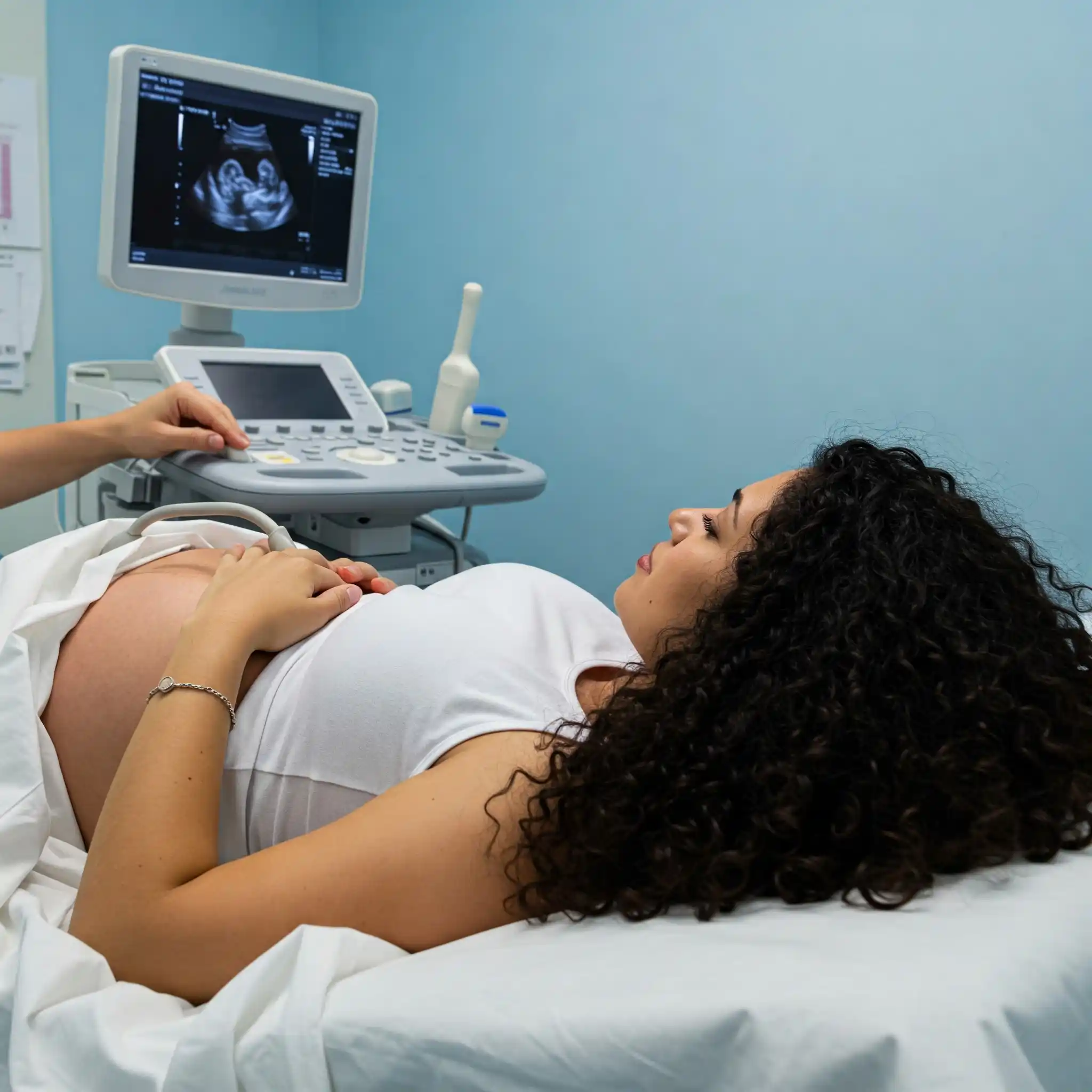
Dating Scan

Also known as the early pregnancy scan, the dating scan is the first scan women need to take after a positive home pregnancy test. This scan is conducted to confirm the pregnancy and establish an estimated due date (EDD) for the baby’s delivery. During this scan, the foetal heart rate is detected for the first time. EDD obtained through this scan is later needed for other prenatal tests conducted in the future. The dating scan is done anywhere between 8-11 weeks of pregnancy.

Nuchal Translucency (NT) Scan

So, what is the nuchal translucency scan in pregnancy? The NT scan is another ultrasound that women take during the first trimester of pregnancy. It is performed between 11-14 weeks and is also done by a licensed sonographer. During this scan, the sonographer measures the fluid-filled space at the back of the foetus’s neck. This measurement is then used to determine the risk of chromosomal abnormalities, like Down’s syndrome and Edwards syndrome, in the developing baby.

2. Second Trimester Pregnancy Scans

Anomaly Scan

An anomaly scan is a comprehensive scan wherein the sonographer examines each part of the baby, right from head to feet. They carefully study the baby’s head, spine, limbs, and important organs like the brain and heart. The aim of this scan is to identify abnormalities, if any, in the foetus. Timely identification helps doctors in deciding the future course of action, such as referring to specialists, if need be.

Cervical Length Scan

This scan is performed between 16-24 weeks of pregnancy and is recommended for women who have previously had late miscarriages or premature labour. It may also be done if a woman shows symptoms of early labour.

The scan is performed by inserting an ultrasound probe into the vagina, where it measures the length of the cervix. Any signs of the cervix shortening could indicate a possibility of preterm labour or early delivery. If the cervical length is short, the doctor then determines the need for further tests or surgical intervention so chances of these issues can be minimised.

Foetal Echocardiography or Foetal Echo

The foetal echo scan is a safe and non-invasive scan performed around 18-24 weeks of pregnancy. It studies the baby’s heart in detail, for example, its structure, functioning, size, etc. It is not advised for all pregnant women and may be required in certain cases, such as:

  • The baby is at risk of having an abnormality in the heart.
  • The baby’s family has a history of heart conditions.
  • The expectant mother has another child who has a heart-related condition.
  • The mother has taken medications or has health conditions that can impact the baby’s heart.

3. Third Trimester Pregnancy Scans

Growth Scan

This scan, which roughly takes around 20-30 minutes, is performed in the third trimester, anywhere between 28-40 weeks of pregnancy. Doctors may advise one or two growth scans, depending on the need.

As the name suggests, a growth scan is done to check the growth of the baby. It shares important details about the baby’s overall health, its position, the amount of amniotic fluid, the position of the placenta and umbilical cord, and more. The scan is also helpful in identifying gestational diabetes, as pregnant women with this condition often reflect higher amounts of amniotic fluid in their bodies.

2D, 3D, and 4D Scans: What’s the Difference?

Besides the scans mentioned above, pregnancy ultrasound scans are also differentiated into 2D scans, 3D scans, and 4D scans.

2D Ultrasound Scan

This is the most basic and commonly used ultrasound scan. Most women have 2D ultrasound scans throughout their pregnancy. These scans produce two-dimensional images, meaning the images look flat and do not have any depth. Despite that, the scan is quite effective in determining foetal growth and health, plus other aspects like multiple pregnancies and ectopic pregnancy.

3D Ultrasound Scan

This scan provides three-dimensional images of the developing baby. The images created are more detailed than those produced by the 2D scan. The test can go on for an hour and is normally done when a woman is 26-30 weeks into pregnancy.

4D Ultrasound Scan

This highly advanced scan is known to offer 3D images of the baby while also allowing you to see the live movements of the baby inside the womb. The scan shows the baby’s face, eyes, swallowing, and other movements. The scan not only offers a treat to the expecting parents, sharing a glimpse of their baby before birth but is also useful in assessing abnormalities that are not very clear in 2D scans.

How to Prepare for a Pregnancy Scan

Pregnancy ultrasounds are usually painless and non-invasive procedures. You will be asked to lie on the examination bed or sit on a reclined couch. The radiologist will then apply a gel on your abdomen and use a device to scan your baby’s growth. Here are a few pointers on how you can prepare for the test:

  • Carry all your doctor's prescriptions, reports, and forms related to your pregnancy. You may not need them, but it is better to carry them if you need them, just in case.
  • Eat normally as you would on any other day.
  • Ask the doctor whether you need a full bladder or an empty bladder for the scan.
  • If you need a full bladder, use the toilet to empty your bladder around 2 hours before the scan. Later, drink at least one full glass of water (or one litre if you can), around an hour before your scheduled appointment.
  • Wear two-piece clothing to make scanning easy and comfortable.
  • Arrive on time so you can relax a bit before the scan.

Summing Up

Ultrasound scans during pregnancy are important for antenatal care. These tests help doctors ascertain whether a pregnancy is proceeding optimally or if there are any issues that need monitoring. By taking timely scans, expecting mothers not only help doctors examine the growth and well-being of their developing baby, but they also get peace of mind, knowing their baby is doing well.

FAQs

What is an ultrasound scan?

An ultrasound scan is a safe, non-invasive, and painless procedure that uses sound waves to create images of the growing foetus. It is commonly used to monitor foetal growth and development.

How many scans will I have during my pregnancy?

The number of scans during pregnancy is not fixed and is usually determined by a doctor. Most women have at least two scans during the pregnancy. However, your doctor may suggest more scans based on your health and that of your baby.

Are ultrasound scans safe for my baby?

Yes, ultrasound scans are safe for the mother and her baby. There is no use of radiation in these scans, and doctors have been using them for several years without a problem.

Can my partner come to the scan?

Yes, your partner or one support person is usually allowed to attend scans with you.

What is the price of an anomaly scan?

Anomaly scan price can range anywhere between Rs. 1,500 and Rs. 5,000 depending on where you are getting it done.


For more health-related content, visit our Dawaa Dost website. You will also get access to quality medicines and healthcare products, all at affordable prices. Check it out now!


Popular Products